Topics in Photographic Preservation 1999, Volume 8, Article 5 (pp. 23-30)
The photography collection of the “Museum for Art and History” in Brussels, Belgium contains a Megalethoscope viewer with 27 sliding plates. On the occasion of an exhibition in 1998, the viewer has been restored. Two master degree students of the Photographic Materials Course of the ‘Royal Academy of Fine Arts of Antwerp’, under survey of their teacher, discussed the conservation of the original plates and made a proposal to the museum staff. All plates were soiled, most plates showed tears in the image- and/or backside. Initially, one plate was considered virtually lost. A plate, in ‘visually good condition’, was chosen and removed from its backmounting to evaluate the ‘inside’ condition. The treatment proposals were based on the inside condition of this plate. This paper will give an overview of the conservation treatment of Megalethoscope plates in varying conditions.
During the 1997–98 and 1998–99 academic years, students of the photographic materials conservation section of the Royal Academy of Fine Arts of Antwerp, Belgium, had the occasion to work on a set of Megalethoscope plates from the collection of the Royal Museum of Art and History in Brussels. The museum staff wanted to have the opportunity to show these original sliding plates in the original Megalethoscope viewer.
The Museum houses an important collection illustrating different civilisations from all over the world. It surveys the history of mankind from prehistoric times to the present, across the five continents. The Museum's task is the collection, preservation and study of evidence of these civilisations, as well as showing them to the public. So, it is both a scientific institution and a museum.
The present form of the Museum is closely associated with the history of its development. The history of the museum begins in 1835, shortly after Belgium gained its independence, but the heart of the collection had been put together much earlier. In 1898, the Belgian government acquired 62 pictorialist photographs from reputed international photographers, which had been exposed at the “IIe Exposition d'Art Photographique de Bruxelles”, and offered them to the “Musée de l'Industrie de Belgique”, which later became the actual “Royal Museum of Art and History”. This purchase was the start of the photographic collection. One hundred years later, in the year 2000, after successive removals and minor disasters, the photographic collection will receive an entire museum wing devoted to photography and moving image.
Carlo Ponti was born in Sagno, Switzerland, between 1822–24. For 8 years, he lived and worked in Paris. Eventually, he settled in Venice where, from 1854 on, he became a successful optician and photographer.1 Renowned for his lenses, Ponti thanked his fame to the invention, in 1861, of his ‘Aletoscopio’, from which the ‘Megaletoscopio’ is derived. Jones2 gives the following description of the Alethoscope:
An optical device invented by Signor Ponti, of Venice, and intended for the inspection of transparencies of ordinary photographic prints. It consisted of a large single lens, suitably mounted… It was claimed that the alethoscope showed single photographs with stereoscopic relief, but that is a theoretical impossibility, although it is possible to obtain a deceptive approximation to relief when looking with both eyes through a large convex lens at a single photograph, provided the lighting, modelling and perspective of the picture are good and natural; but this effect is more due to imagination and suggestion than anything else.…
During the 1860's, Ponti published a series of photographic albums under the title “Ricordi di Venezia”. He also published and edited photographs from other Venetian photographers. The administrative confusion occasioned by the Austrio-Prussian war and the following Peace Treaty of 1860 caused Carlo Ponti to loose the copyright on the alethoscope. His collaborator, Carlo Naya, began producing the Alethoscope under his own name. As long as he was professionally active he tried, without success, to regain his privilege. Carlo Ponti died, completely blind, in 1893.
The megalethoscope is an enlarged version of the alethoscope and confides its confection to the cabinet-maker Demetrio Puppolin, whose name also figures on different megalethoscopes. A curved viewing slide was placed in the instrument, at the rear. The observer looked into the show box through the eye-glass of the arrangement. The scene could be viewed by reflected or transmitted light by manipulating the hinged top and side doors.
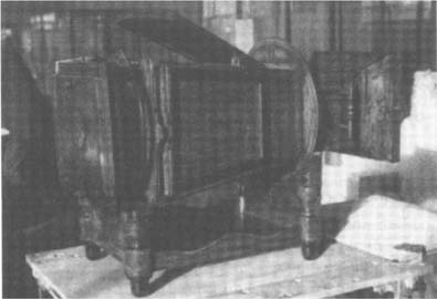
These instruments existed in different versions: in simple walnut; with inlaid-work; or with sculptured adornments. An exceptional version was called the ‘Megaletoscopio privilegiato’, it was designed to receive curved plates, of which the Museum of Art and History has a copy.
Figure 1: ‘Megalethoscopio priviligiato’.
The plates for this version of the viewer are curved. We are not sure why, but our opinion is that Ponti would obtain an optical correction of lens aberrations. The plates were constructed for viewing photographic images under reflected and transmitted light to suggest day or night lighting. All photographic prints on these plates were albumen prints. Pinprick perforations and mechanical thinned areas on the albumen prints have been made to create different light effects. The pine wooden strainers were made by using ‘channel groove’ join attachments with iron nails. On the short sides of the plates, wooden handles were attached for handling. The strainer was painted black, included the front protective paper borders. Some of the plates were provide with printed labels describing the image subject. The strainer measures about 30 × 40 cm, the image about 24 × 32 cm. The cross section of a plate ( fig 2) shows two or (mostly) three layers:
Figure 2: Cross section of a megalethoscope plate
All plates were extremely soiled by dust, some by diverse adhesions. Half of the plates showed small tears in the primary and secondary supports. Twelve plates showed major tears in the photographic print. One plate was considered virtually lost because of a great number of tears and a gap in the print. All secondary supports showed brown spots. Some plates had lost one or both handles.
In order to make further decisions, a plate in relative visual good condition was first dry cleaned and removed from its secondary support to evaluate the inside condition. The back paper of the primary support had been painted with watercolour and was in good condition. Opposite the pinpricks, on the paper side of the secondary support, were some small soiled areas. There were no traces of biological deterioration. The paper of the secondary support was brittle and discoloured. Measuring of the transmission density of the secondary support resulted in a value of approximately 0.80.
This first plate did not contain an intermediate paper but by removing the secondary supports of the other plates, we could assess that most of the plates had been mounted with a blue tissue or white waxed paper between the primary and secondary support.
Most of the blue tissue papers were torn, very brittle, and were discoloured on the borders (due to contact with the wood of the strainer).
A treatment scenario was considered that made the plates useful in the least intrusive way. Removal of the secondary support would give easy access to the back of the primary support in order to repair the tears in the photographic prints. The primary support would remain on the strainer. There was no need renewing the textile layer of the primary support; the risk of damaging the photographic print and/or the watercolour painted paper was considered too high. The secondary support would be replaced by a more transparent paper/textile layer; the objective was a density value between 0.40 and 0.60. After dry and wet cleaning, repairing the tears, and retouching the repairs, a new secondary support and paper border protections would be mounted. The extant labels would be replaced.
First, the condition of each plate was drawn up. After dry and wet cleaning (ethanol/demin, water, 30/70) of the albumen print, the secondary support was removed by using a firm (strong) starch glue. The paper border protection could not be recuperated. The interior was been dry cleaned using a soft brush. The tears in the primary support were repaired from the back by using starch glue, and drying under smooth pressure between synthetic supports. The handcolouring along the consolidated tears has been retouched.
The fragile intermediate blue tissue papers must be lined before remounting them. This was done by using a Bolloré paper, and was done at the same time the tears were repaired. The repairs were retouched by using watercolour paint using transmitted from a light table.
We evaluated different paper/textile combinations on their shrinking capacities, and transmission density. We choose three, more or less similar to the original, different, pure cotton textiles and lined them with the Bollore paper. Bolloré paper is a strong Western, type quality paper with well-distributed fibres, and has a (for this purpose) good transmission density (around 0.20), and is less expensive than the most Japanese papers.
The textiles were washed in hot water to remove additional products such as starch (apret). After lining they were immediately wet mounted on a wooden frame. The least shrunken, combination was selected for further use, but first we did another test. The selected textile had a different number of chain (10) and woof (18) wires, which resulted in different shrinking behaviour by using the textile in different directions on the plate. Mounted with the weft-side parallel to the long side of the plate, after drying, the secondary support could make contact with the primary support. Mounted with the warp-side parallel to the long side of the plate, there was less shrinking in the long side direction and the supports could not make contact. This was the best way of mounting. For manipulating the new secondary support, the size of the textile and paper should be at least 20 cm larger on both sides so it can be pinned to a board.
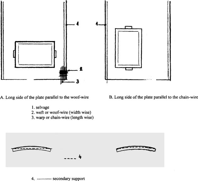
Figure 3: Shrinking behaviour of the new textile/paper combination
For the newly lined secondary support, we measured a transmission density of approximately 0.40. In combination with the lined blue tissue this resulted in a total density of approximately 0.65.
We divided the plates into two groups: plates without and plates with an intermediate paper. For plates without an intermediate paper, mounting of the secondary support could be executed wet in one step. For plates with an intermediate paper, mounting was executed in two, dry, steps. The secondary support was lined and dried in advance. Then, the lined intermediate paper was adhered to the borders by using starch glue. The near dry secondary support was mounted the same way and stretched on the strainer.
The treatment of this plate required complete dismantling. Both the primary and secondary textile layers were replaced. The albumen print and the watercolour painted paper were adhered, by using a firm (strong) starch glue, on the new textile layer. Tears were filled by using paper fibres. The gap was repaired by filling it with a similar paper and retouching the paper by using watercolours.
A list of the treatment steps:
Mounting:
The first objective was to find a minimally intrusive treatment; it was not necessary to remove all the albumen prints from their textile support. Replacing the secondary support was justified, because both the paper and textile layer were soiled and damaged and the paper was brittle. The pH of the paper and textile was tested, resulting in values of approximately 4.9. This case study has given us the opportunity to understand these types of objects, especially the treatment of the “virtually lost” plate.
We wish to thank our teacher Roger Kockaerts for his support, and Madame Deltour of the Museum of Art and History for giving us the opportunity to work with these objects and the PMG specialty group for the possibility to come into the conservators' world.
BALZER RICHARD, 1987, “Optical amusements: Magic lanterns and other transforming images”, Mass., USA.
CARLO PONTI, 1997, “Un magicien de l'image”, Exposition catalogue, Exposition au musée Suisse de l'appareil photographique Vevey, du 31 octobre 1996 au 2 février 1997.
DUNE CORINNE, 1996, “La photographie en spectacle, traitements de papiers albuminés peints”, Coré n° 1, septembre 1996.
GASCOIGNE BAMBER, 1995, first ed. 1986, “How to identify prints, a complete guide to manual and mechanical processes from woodcut to ink-jet”, Thames and Hudson, London.
HENISCH, Heinz & Bridget, 1996, ‘The painted photograph 1839–1914’, Pennsylvania Press.
MANNONI, Laurent, 1996, ‘Le mouvement continué’, catalogue illustré de la collection des appareils de la Cinémathèque, Paris.
STANLEY, TED, 1996, “A Conservation case study of polyrama panoptique paper viewing slides”, JAIC 35, 1996.
1 Musée Suisse de l'appareil photographique, 1996, ‘Carlo Ponti — Un magicien de l'image’, Vevey
2 JONES, E. Bernard, 1911, Reprint 1981, ‘The Encyclopaedia of Early Photography’, London, Reprint from: Encyclopaedia of Photography.