Topics in Photographic Preservation 2009, Volume 13, Article 17 (pp. 110-126)
Presented at the 2009 PMG Winter Meeting in Tucson, Arizona
Platinum, palladium, gum dichromate, gelatin silver, and photogravure are among the processes found in the collection of Alfred Stieglitz. His work is a testament to both the aesthetic and technical evolution of American photography, from the end of the 19th to the middle of the 20th century. His early work was without restriction -he used many techniques and experimented with a vast array of materials. Over time, he moved away from hand-made photographs toward more commercially-produced photographic papers. This evolution is not only visible in his work, but also in the works he collected during his life. After his death in 1946, his widow, Georgia O’Keeffe, divided and donated the collection to several institutions, including a major gift to the Art Institute of Chicago, but process identifications were sometimes absent or misleading. There have been limited attempts by the Art Institute of Chicago in the past to accurately identify certain photographs in the collection, but due to limitations of time and equipment, no comprehensive effort toward this goal has been undertaken. In the current project a protocol is developed for the analytical equipment at the Art Institute to map out the Alfred Stieglitz collection.
Keywords: photography, photographic techniques: FITR-(ATR); Raman; FT-Raman; XRF; Alfred Stieglitz
Museum professionals, experts and collectors typically rely on visual examination to identify photographic processes. However, visual identification alone can be misleading. The present study describes applications of several instrumental analyses used to identify organic and inorganic materials non-destructively.
A protocol was developed for two types of X-ray fluorescence spectrometers, a portable micro-focus system and a handheld macro-set-up, and validated with standard replicas of known photographic techniques. Qualitative XRF identification of the metallic salts allows unambiguous identification of the process used for each photograph.
The presence of a coating complicates the identification of the used medium. Therefore a protocol on non-destructive analyses (Infrared and Raman Spectrometry) for coatings was developed. The study investigated several combinations of coatings on photographs. For the comparisons with unknown coatings three types of photographic paper (one, two and three layered papers) and five primary components of coatings were used to develop different model systems. The next step was to use these models in case studies and is now being used to identify photographs in the Alfred Stieglitz collection.
With photography increasingly recognized as a distinct artistic medium, the Alfred Stieglitz (1864-1946) Collection has gained great historical significance, and is now one of the most important collections of photographs at the Art Institute of Chicago. In addition to creating his own work, Stieglitz also discovered, promoted and collected the work of many other photographers, wrote articles, and published magazines – all with a singular goal: the recognition of photography as an independent art form. The breadth and quality of the work he had amassed by the end of his life set a precedent for subsequent collections of photography. His collection represents an artist who explored the photographic medium to find the techniques and materials that would reflect his ideas and, rather than providing an historical overview of a given artist, speaks to the interest and collaboration of Alfred Stieglitz with that particular person. This collection was divided and donated to several museums, archives and universities. According to Georgia O’Keefe, Stieglitz’s widow, this was the ideal way to show the work to the largest possible audience.[1]
The Alfred Stieglitz collection captures a defining moment for the creation of visual vocabulary of modern photograph. There is a great variety in techniques present in the collection: autochrome, carbon, gelatin silver, gum dichromate, photogravure, platinum, palladium, Satista, salt print and combinations thereof.
It is useful to categorize photographs as having one, two, or three layers. Within these categories, aspects such as size, color and finishing are important parameters for the estimation of the technique used. It is often possible to distinguish photographic techniques by visual identification, relying on individual characteristics such as grain pattern, layer structure and other special features.[2] However, artists have access to a great number of different products and options to translate their ideas into photographic imagery resulting in a melting pot of both commercially produced photopapers and manipulated handmade photographs. Stieglitz was widely known to experiment with different sensitizers, developers, and toners to achieve a specific aesthetic result, which can further complicate the identification procedure.[3]
The artist also strongly believed in the presentation of a photograph, which often-included mounting the photograph on a specific paper and applying a coating on the image surface. He used coatings for both aesthetic reasons as well as preservation enhancements.[4] The application and type of coating used varies from minimal to one drastically altering the surface and perception of a photograph.[5] The presence of a coating layer can also interfere with accurate visual identification. There is an abundance of materials and application methods that can be used.
The composition of metallic elements is directly related to the used technique and the structure of the photopaper. Salt prints, platinum, Satista, palladium and platinum-palladium are the most common single-layered photographs in the Alfred Stieglitz Collection. One-layered photographs have a matte surface, no binder or baryta layer, and the paper fibers are clearly visible. These techniques can sometimes be distinguished by the color of the print. In a salt print Ag and Cl make up the image material, while Au can be present as a toner. In contrast to the salt print, the platinum or palladium process is based on the light sensitivity of iron salts, which react with the platinum or palladium compounds to produce a non-silver image.[6] Platinum prints often have a neutral grey-black hue while palladium prints tend to have a warmer brown tone. The two processes have very similar chemical reactions – they are often interchangeable. In a platinum print Fe, Pt, is used for the image with possible toning, development or sensitization achieved with U, Hg, and Au. In a palladium print, almost the same elements are found but pt is replaced with pd. When a combination of the two salts is used both pt and pd are present in the final image. In the Satista process the characteristic elements are Pt, Ag and Fe.
In photographs with two layers, there is a binder present but not a baryta layer, so the paper fibers remain visible in the highlights. The Stieglitz Collection contains carbon and gum dichromate prints. Both techniques are based on the light sensitive properties of dichromate slats and fine pigments suspended in a binder such as gum arabic for the gum dichromate prints and gelatin for carbon prints. During the Pictorialist movement this technique was often used in combination with other techniques. It is difficult to distinguish between carbon and gum dichromate with XRF, as both have the same characteristic elements: Cr, Fe and pigments.
In photographs with three layers, there is a binder and a separate baryta layer, which obscures the paper fibers.[3] The binder may be gelatin or collodion, in which a silver emulsion has been suspended. The baryta layer is a combination of BaSO4 and gelatin.
X-ray fluorescence spectrometry (XRF) is a non-destructive analytical tool often used in photograph conservation to identify the elemental composition of a photograph. XRF is based on the ionization of atoms of the material being investigated by an energetic beam of primary X-rays. The characteristic radiation or fluorescence X-ray that is emitted by the ionized atoms contains information on the nature of the present elements. In the past decade, several publications have described applications of XRF to art analysis.[7-13] The elements of portability, non-contact, characterization through multi-elemental fingerprinting, and both qualitative and quantitative analysis capabilities are very attractive features for cultural heritage applications.
Previous applications of XRF to the study of photographs include identification of photographic techniques to support art historical research, the quantification of BaSO4 for authentication and dating of photographs and the examination of differences between traditional developed gelatin silver prints and developer incorporated stabilized photographs.[3,15] The work described here builds on the foundations established by previous studies conducted at The National Gallery of Art in Washington, DC on the use of XRF spectrometry to characterize the Gallery’s collection of Stieglitz prints.[3,16] Since the time of those publications many improvements in portable and handheld X-ray fluorescence instrumentation made it possible to better detection of the elements that characterize the final tone and technique of the photographs, without the need for tailored secondary target excitation.
Identification of the photographs is a two-part process. It is possible to detect the metallic elements with XRF but questions on the organic compounds are not answered. In several cases the binding materials are as important to identify the technique. A good example is the presence of Cr; it can be a gum dichromate or a carbon print. More important is the presence of a coating. As coatings are the top layer of the photograph, their condition and identification is important.
The coatings may degrade or get damaged, critically affecting the photograph and the perception of the image. Thus, sensitive detection and accurate identification of a photographic coating, together with the determination of its condition, is crucial to inform appropriate conservation treatments. Among non-invasive techniques of analysis, FTIR-ATR has been the method of choice for the identification of coatings on photographs, whereas little attention has been devoted to the study of these films with Raman spectroscopy.[17] Fourier transform Raman spectrometry measures the wavelength and intensity of inelastic scattered light from molecules while using an interferometer and a 1064 nm laser source. The use of the 1064nm source eliminates the fluorescence of the samples. The Raman technique has the advantage of being not only non-invasive, but also non–contact, overcoming one shortcoming of FTIR-ATR, namely a residual small indentation left on the surface of the artwork as a result of the analysis.
Laboratory simulations allowed the determination of the thermal sensitivity threshold of each combination, correlation of coating thickness, as well as detection capabilities and limitations. FT-Raman was compared to other complimentary vibrational spectroscopy techniques such as micro-FTIR-ATR and micro-FTIR performed in transmission with diamond anvil cell.
XRF analyses
Bruker ArtTAX air-path portable μ-XRF system with spot size of 0.2 to 2 mm, micrometric control of the position of the measuring head with respect to sample, laser pointer and integrated camera system that allow optimal positioning at a fixed distance of approximately 5 mm from the sample surface. The system is equipped with interchangeable excitation tubes with Mo and W targets. (max. 50 kV, max. 800 μA) and 0.2 mm air cooled Be window with Cu-radiator. The X-ray detector is an X-Flash® detector (5 mm2) with energy resolution of 165 eV for the full width at half maximum of the MnKáline at 10kcps
Bruker/KeymasterTRACeR III-VTM energy dispersive handheld X ray Fluorescence analyzer, with Peltier cooled advanced high-resolution Silver-free SiPIN detector with a 13μm Be window and resolution of approximately 175 eV for the full width at half maximum of the MnKáline. The system has titanium and aluminum changeable filters, and is equipped with a rhodium (Rh) transmission target with max voltage of 45kV and tunable beam current of 2-25μA.
FT-Raman analyses
A Bruker Vertex 70 FTIR Spectrometer coupled with Ram II FT-Raman module and Ramscope III FT-Raman microscope was used. The system is equipped with Nd3+/YAG laser, with excitation at 1064 nm and high sensitivity, nitrogen cooled Ge detector. Between 5000-50 000 scans were accumulated at 4 cm-1 resolution using a 10x objective, with a nominal laser power of 5-500 mW.
FTIR analyses
FTIR spectrophotometer a Bruker tensor 27 FTIR spectrophotometer with mid-IR glowbar source and DTGS detector was used, coupled to Hyperion 2000 Automated FTIR microscope with nitrogen cooled mid-band and broad-band MCT detectors (covering the range 7000-). Sampling accessories with ATR objective (germanium crystal) and diamond micro compression cell in connection with microscope at a resolution of 4 cm-1 for 256 seconds.
Detection of metallic elements with XRF*
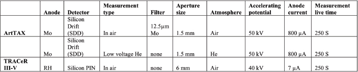
Different conditions with both instruments on facsimiles developed at the National Gallery, Washington DC. were used to validate a protocol.[3, 18] In table 1 an overview of the different settings is given. If radiation dose is a concern, low KV settings in conjunction with He flushing and evaluation of the M lines for Pt and Pd and L lines for Pd Ag Hg and Au is a possibility. However, peak overlap is more pronounced and the sensitivity lower than when operating at higher voltages and with primary beam filtration, which provided optimal conditions for detection of the metallic elements.
Table 1: Experimental conditions used for the XRF analysis of the Stieglitz collection with portable(ArtTAX) and handheld (TRACeR III-V) spectrometers
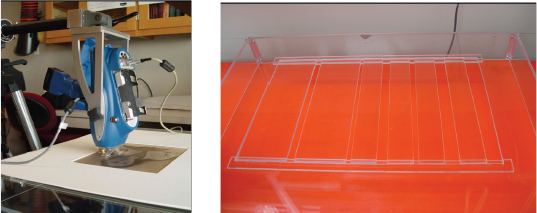
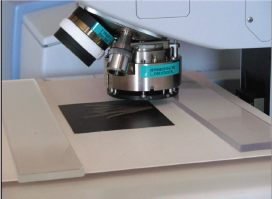
To reduce the background of the spectrum and improving the signal-to-noise ratio a acrylic stage was specially designed The photographs were placed flat on a specially designed acrylic stage, the stage has a adaptable support bars allowing the opening to change to support the photograph were necessary and enables sampling in air (Figs. 1, 2). The horizontal acrylic stage is advantageous for the analysis, poses less stress and strain on the artifact. All analyses were performed in no-contact mode, for the Keymaster TRACeR III-V the instrument is placed perpendicular to the surface and is supported by a tripod over the acrylic stage, hovering at a 2-3 mm distance from the surface (Fig. 1). The ArtTAX measuring head is angled at 45 degrees towards the photograph to minimize scattering and is separated from its surface by about 5 mm.
Figure 1: Experimental set-up with handheld TRACeR III-V positioned over portrait of Georgia O’Keefe (AIC 1949.745A) by Alfred Stieglitz
Figure 2: Acrylic stage for analysis of photographs and other works of art on paper
The use of XRF in characterizing photographs brings a unique set of challenges to data interpretation. Due to the low amounts of metallic elements in a thin layer, even with long acquisition times, there are detection limitations from the technique as well as extraneous peaks deriving from instrumental contaminations in our case Ni, Cu and Zn are present inside the instrument. Additionally one has to be aware that the collected information is not only related to the image material but also to its paper and its support.
To overcome matrix and instrumental interference and to guarantee macroscopic homogeneity of samples, several analytical spots were selected on each artwork. This resulted in multiple spectra taken from different density areas, as well as spectra of the different paper layers and museum mounting board. By collecting the spectra of each component of the mounted photographs, and overlaying the data it is possible to clearly identify the elemental peaks that are solely related to the image material.
Development of protocol for non-destructive FT-Raman spectroscopy on photographs
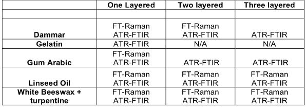
The study used four types of photographic papers (salted paper, cyanotype, albumen and gelatin silver paper) and five primary components of coatings (dammar, gelatin, gum arabic, linseed oil and white beeswax used independently or diluted with turpentine). On the photographic paper, step wedges were printed to test the entire range of densities of the photographs. To compare the coated photographs, bulk samples as well as photo papers without coatings were prepared and analyzed with the same analytical techniques.
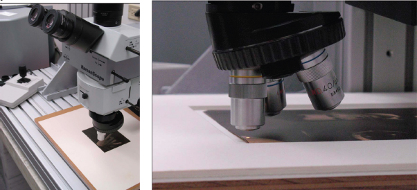
The Ramanscope III FT-Raman microscope can be mounted to a metallic stage that allows to analysis of photographs without removing them from the mounts or restriction in the location of the analysis.
Figure 3: FT-Raman set-up with photograph by Alfred Stiegltiz
Figure 4: Detail FT-Raman set-up with Self-portrait of Edward Steichen
Identification of coatings on different photographic papers
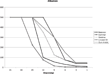
To avoid thermal damage and possible degradation of the sample the maximum power for each type of coating and paper was determined. At the minimum density (step 21) maximum nominal power (at 500 mW) showed no thermal damage. The density of the paper affects thermal sensitivity and the darker the area the lower the power that can be used, due to enhanced absorption of the laser light. There is an inverse relationship between the density of the sample and the maximum power that can be used for analysis. It was also clear that each combination of photo paper and coating has different thermal damage thresholds. There is a tendency for the one layered papers to be more sensitive to thermal damage than the two or three-layered papers.
Figure 5: Power that can be used before thermal damage
Detection limits with FT-Raman[19]
The thickness of the coating affects the detection limit of the FT-Raman. To have an idea of the detection limits in terms of coating thickness a semi-quantitative study was carried out. White beeswax and turpentine were applied in three thicknesses (0.5 mm, 0.25mm and 0.05mm) on salted paper. This resulted in actual coatings measured on SEM images of 1.5μm, 2.5 μm and 150 μm. The FT-Raman spectra were collected at 50 mW with a 10x objective for 20 000 scans. The ratios of the relative intensities of the Raman bands corresponding to the beeswax and the paper were used for data evaluation for this semi-quantitative study. The ratio intensities at 2848 cm-1 and 1379 cm-1 for white beeswax and paper were used respectively. It can be concluded that thicknesses > 3μm coatings can be detected with the FT-Raman.
Detection capabilities of the FT-Raman[20-23]
The Raman spectra were recorded for 5000-50000 scans at 4cm-1 resolution with a nominal laser power of 50-200 mW (resulting in actual powers measured at the sample of 25 – 100 mW). The laser was focused on the coatings of the different photographs, using a 10x objective. With the 10x objective more light is back collected into microscope allowing for a increased signal.
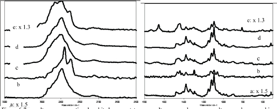
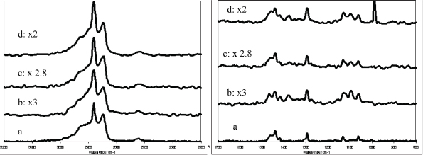
Figure 6-7: a: salt paper no coating, b: white beeswax+ turp. on salt paper, c: dammar on salt paper, d: gum arabic on salt paper and e: linseed oil on salt paper
In figure 6 and 7 the different coatings, detected by the FT-Raman, on salt paper are presented. Although peaks related to the paper substrate are still visible in the spectra, the peaks of the applied organic materials are still distinguished and characteristic so that the coating can be identified.
Dammar (fig 6-7) as a coating was detected with the FT-Raman on salted paper, cyanotype and albumen but not on gelatin silver paper (POP) possibly due to the thinness of the coating on this non-absorptive paper. In the region between 3000 and 2800 cm-1 in addition to the peak around 2894 cm-1 characteristic of the paper, a band at 2922 cm-1 frequency is visible characteristic of CH stretchings of the Dammar. Also the peaks at 1450 and 712 cm-1 are more pronounced when compared to reference spectra of uncoated papers. It is sometimes challenging to compare unprocessed spectra of coated and uncoated papers, but spectral subtraction of reference spectrum of the paper substrate from the spectrum of the coated papers allows to enhance the peaks due to the coating only.
Linseed oil (fig 6-7) as a coating is detected with the FT-Raman on one-layered papers (salted paper, cyanotype), two-layered paper (albumen) and three-layered papers (POP paper). Peaks at 1744, 1439, 1070, 800 and 600 cm-1 were detected with the FT-Raman for linseed oil can be distinguished from the peaks of the paper.
Several of the peaks of gum arabic (fig 6-7) overlap with the peaks of the paper. To evaluate the coating of gum arabic a spectral subtraction provides a clearer identification. Peaks at 1461, 1340, 1261, 979, 879, 842 cm-1 were detected after subtraction of the paper. FT-Raman detected the gum arabic on one-layered photographs but not on the two-layered and three-layered photographs.
FT-Raman does not detect gelatin on one, two, or three-layered photographs.
Pure white beeswax and white beeswax diluted with turpentine are detected with the FT-Raman on the one-layered, two-layered and three-layered papers.
Figure 8-9: a: white beeswax (bulk), b: white beeswax on salt paper, c: white beeswax on albumen, d: white beeswax on gelatin silver paper (pop)
In the region between 3000 and 2800 cm-1 clearly visible and the sharp absorption at 2922 cm-1 typical of white beeswax. Also the peaks at 1439, 1294 and 890 cm-1 are evident, superimposed to the spectral signature of the paper substrate.
Comparison of the non-contact FT-Raman technique with ATR-FTIR and FTIR
FTIR-ATR is a widely used analytical technique to identify films and coatings compounds without sampling. The goal of these test was to see where the two techniques in parallel to each other to gain the best information of the coatings.[24] As FT-Raman cannot detect all the different materials the use of FTIR-ATR can complete the gathered information. Samples were also analyzed in transmission through the microscope after compression in a diamond micro-compression cell.
Figure 10: Set up ATR-FTIR
The analyses with the FTIR-ATR were on the same samples as the FT-Raman. The FTIR- ATR detected all the different coatings on the one, two, and three-layered photographs with the exception of the gelatin coating on albumen and pop paper. Infrared spectrometry cannot make a distinction between proteinacous materials of different origin but only the general class of compounds is identified. The ATR-objective is pressed onto the coatings of the photo papers. The advantage of the ATR-objective is that it only penetrates a few micrometers so there is less interference of the paper as compared with the FT-Raman. One drawback of the FTIR-ATR is that it leaves a residual small indentation on the surface of the artwork as a result of the analysis. The ATR-objective leaves a larger indentation on the three-layered photographs. Depending of the type of coating the diamond leaves a circular indentation with possible cracking (fig 13). On the other hand, with one layered papers the fibers can relax back after contact with the ATR crystal and so the analysis leaves virtually no trace (fig 14).
Figure 11 and 12: Indentation of ATR- objective on a pop paper with a linseed oil coating (left), Indentation of ATR-objective on a pop paper with a gum arabic coating (right)
Figure 13 and 14 : Indentation of ATR- objective on a pop paper with a dammar coating (left), Indentation of ATR-objective on a salt paper with a dammar coating (right)
In case sampling is an option transmission infrared spectrometry can be applied. With the diamond cell the different compounds can be identified without interference of the paper.
Only a small amount, a few micrometers, is required to perform the analysis. The sampling is not visible with the naked eye, unless there is an extremely thin coating.[25]
Overview of non-destructive techniques that can be used to identify coatings
Results and discussion
In total 62 out of 250 photographs of the Alfred Stieglitz Collection were tested with XRF. The selection within the collection was made based on the uncommon appearance of the photographs, questionable information on earlier records, and specific requests by other researchers.
The XRF analyses clearly identified all the characteristic elements for the photographs.
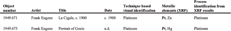
Attributions of the various elements to specific steps in the process of creating the final image were hypothesized, based on the total area of the element’s peaks detected in the high density, low density or paper substrates of the photographs. In total, 36 platinum prints were identified, 17 palladium, 3 gum dichromate, 2 gum dichromate over platinum, 1 gelatin silver and 1 platinum-palladium. Table 2 gives an overview of the photographs that have been tested.
The FT-Raman detects drying oils, waxes, resinous media and gums on photograph papers (albumen, cyanotype, salted paper, and pop paper) only when thickness is > 3 μm. Due to the interference of the paper the coating has to be thicker than 3 μm to be detected. The best results were gathered on one-layered papers and especially on salted paper. To obtain the spectra long collection times are necessary. There is interference from the paper substrate that makes data collection and interpretation laborious. In the following, specific examples are discussed regarding photographs whose technique presented challenges to visual identification that were unraveled by instrumental analysis.
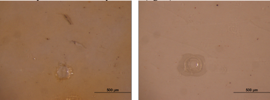
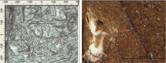
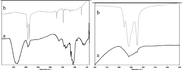
A portrait of Georgia Engelhard (1949.716) by Alfred Stieglitz was identified as a palladium print however micrographs showed a glossy surface. XRF analysis with the ArtTAX confirmed that pd was the image material and that the glossy surface there is most likely a coating. Due to the thinness of the layer initial measurements with the FT-Raman did not identify the coating. Therefore a small sample (a few microns) was taken from a damaged are in the corner outside the image. The coating was identified with diamond cell FTIR as an extremely thin wax coating.
Figure 15: Micrograph of surface of Portrait (1949.716) by Alfred Stieglitz
Figure 16: Micrograph after sampling, he white paper fibers are evident fro a pre-existing area of loss
Figure 17-18: Spectrum a is taken from the is the photograph with coating compared to spectrum b, a bulk samples of beeswax. In spectrum a from the photograph, contributions from the beeswax coating are evident, superimposed to the paper absorption and are especially visible in the range 3000- 2800 and 730- 720 cm-1. On the right a detail of the CH stretching with b reference beeswax and a the coated paper fiber.
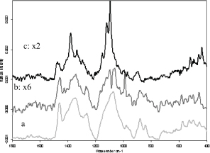
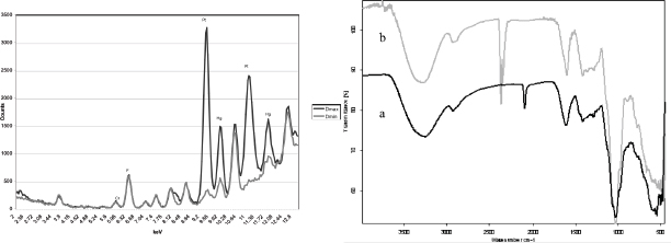
A photograph of Georgia O’Keeffe’s hands (1949.743) by Alfred Stieglitz was visually identified as a palladium print. Under magnification there was a atypical hue and cracks evident indication that a coating was present. XRF analysis with the handheld spectrometer confirmed that the image is produced with Pd and also contains Hg. Hg can be present in the sensitizer, developer or toner and is responsible for the warmer hue of the print. Data was collected with the FT-Raman for 8000 scans at 100 mW. When comparing it to reference samples of coated salt papers the coating can be identified as a gum Arabic. There are still some questions that the coating maybe a mixture of gum and wax. Longer acquisitions will provide more accurate data.
Figure 20: Spectra a is the bulk of Gum Arabic, spectra b is the subtraction of gum Arabic (reference) on salt paper (reference) and c is the spectra from the photograph by Alfred Stieglitz
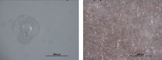
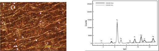
A portrait of Georgia O’Keeffe (1949.745A AIC) by Alfred Stieglitz was described in the past as a gelatin silver print with yellowing at the edges. In 1989 the portrait of Georgia O’Keeffe was identified with XRF as a palladium print with traces of platinum and gold. The micrograph see fig. 22n clearly shows cracks and a substance on the surface of the palladium print.
Figure 21: Micrograph of Georgia O’Keeffe hands (1949.743 AIC)
Figure 22: Spectra a is the reference of wax on salted paper, spectra b is the spectra of the photograph of Georgia O’Keeffe hands
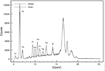
Girl with a muff (1949.856) by Clarence H. White, visually identified as a Pt print, was examined because other prints of this same negative exist and the technique was questioned. XRF proved the presence of pt in the image. However, analysis also detected the presence of Hg, which could explain the warm tone of the image, a characteristic typically not associated with Pt prints.
Figure 23: XRF results for the photograph of Girl with a muff (1949.856 AIC). The difference between the max and min density is clearly visible, pt and hg are present in larger quantities in the darker area’s indicating that they are used as medium for the image.
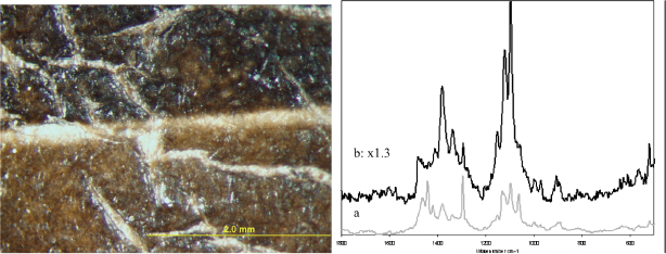
A portrait of Clarence White by Edward Steichen is (1979.828) was in the past identified as a platinum print with mercury toning. The visual identification showed a very glossy surface with cracks (Fig. 23). XRF identified the elements, Pt, Hg, Fe and Cr. The presence of Pt and Hg suggest a platinum print, and the chromium points to a gum dichromate layer. The top layer of the photograph was later indentified with ATR-FTIR as a gum Arabic. The photograph of Clarence White can with certainty be identified as a gum dichromate print on top of platinum with mercury print.
Figure 24: micrograph of the portrait of Clarence White by Edward Steichen is (1979.828 AIC), the cracks are clearly visible. Figure 25 is XRF spectra of the max and min density with the ArtTAX[18]
The photograph Moonrise (1949.830) made by Steichen at Lake George is an excellent example of how the combination of analytical data with archival research can lead to an enhanced understanding of the photographic processes. In the past this photograph was associated with several different techniques. Two proposed identifications suggested the combined technique of gum over cyanotype or gum over platinum print. The XRF spectrum illustrated in Fig. 25 shows the presence of Pt, Cr, Fe and Zn, so it can be said with certainty that this is a platinum print. On the other hand the element chromium indicates a gum dichromate print, with an iron-based pigment. The ATR-FTIR results show clearly that the top layer is gum Arabic and that the iron based pigment is Prussian blue.[26]
This knowledge, supplemented by correspondence between Alfred Stieglitz and Edward Steichen can allow identification of the photograph as 3 prints: From a letter by Edward Steichen to Alfred Stieglitz on the technique of the photograph: “Moonrise: 16x 24[1904] in three printings: first printing, grey black platinum, 2nd, plain blue print (secret) and 3rd greenish gum”.
Figure 26: XRF results of Edward Steichen’s Moonrise
Figure 27: Spectra b is the reference sample of gum arabic with ATR-FTIR and spectra a is the Moonrise (1949.830 AIC) of Edward Steichen
The goal of the project was to develop a protocol to characterize photographs by identifying the elements used in the process and organic components presents in the binder or as coatings by fingerprinting the materials with vibrational spectroscopic techniques. Two different XRF instruments were used to develop a protocol enabling a clear identification of the characteristic elements. As a result, we proved that satisfactory process identification can be achieved both with a laboratory set-up, but also with a hand-held system that can be carried in storage spaces, conservation labs or galleries with ease. To identify organic compounds a second protocol was developed with special attention to the non-destructive, non-contact FT-Raman spectroscopy technique. FT-Raman proved promising results for positive identification of coatings on various systems, but has limitations in its ability to identify all organic compounds especially when the films are very thin. Also the collection of the data is a long process. FTIR-ATR is faster in collecting the data, is able to detect all the most common traditional coatings materials but leaves an indentation on the surface, which is especially visible in three layered photographs. Also there is a limitation on the location of the analysis because of the FTIR-ATR set-up. There is a limitation on the width of the mount that allows the crystal to still make contact with the actual image. Alternatively, by taking a small sample of a damaged area or corner the coating can be analyzed with the diamond cell in transmission FTIR. FTIR is a quick effective technique to analyze a coating without the interference of the substrate. At the moment this protocol is being used at the Art Institute of Chicago to identify organic and inorganic compounds on photographs. Depending on the tools and the object, different analytical approaches can be used to identify the organic compounds. At the conclusion of the project, among the 62 tested photographs, 8 that had been incorrectly identified were rectified, 13 more although correctly identified by visual observation gained additional fabrication information as a result of the XRF study, and 26 analyses confirmed the existing medium display, allowing the museum to remove long-standing question marks on exhibition labels and media description information.
[1] O’Keeffe, G. 1949. Stieglitz: His Pictures Collected Him. New York Times: 24.
[2] Reilly, M. J. 1986. Identification of 19th Century photographs. Kodak, New York: 48.
[3] McCabe, C. and L. D. Glinsman. 1995. Understanding Alfred Stieglitz’ Platinum and Palladium prints: Examination by X-ray fluorescence Spectrometry. Conservation Research, NGA, Wash. D.C.: 73.
[4] McCabe, C. 2005. Coatings on photographs by Alfred Stieglitz. Coatings on Photographs Materials Techniques and Conservation, (Ed. C. McCabe), PMG-AIC, Wash. D.C.: 301-313.
[5] Von Waldthausen, C. 2005. Coatings on Salted Paper, Albumen, And Platinum Prints. Coatings on Photographs Materials Techniques and Conservation, (Ed. C.McCabe), PMG-AIC, Wash. D.C.: 79.
[6] Crawford, W. 1979. The keepers of Light. A history & working guide to early photographic processes. Morgan & Morgan , New York: 167.
[7] Moioli, P. and C. Seccaroni. 2000. Analysis of art objects using a portable x-ray fluorescence spectrometer. X-Ray Spectrom 29: 48.
[8] Janssens, K. G. Vittiglio et al. 2000. Use of Microscopic XRF for Non-destructive Analysis in Art and Archaeometry. X-Ray Spectrom 29: 73.
[9] Janssens, K. 2004. Comprehensive Analytical Chemistry, Vol XLII Non destructive Microanalysis of Cultural Heritage. Elsevier B. V. Amsterdam, The Netherlands.
[11] Caneva, C and M. Ferretti. 2000. XRF Spectrometers for non-destructive investigations in art and archaeology: the cost of portability. 15th World Conference on Non-Destructive Testing (http://www.ndt.net/article/wcndt00/papers/idn680/idn680.htm accessed on 2/27/09).
[12] Scott.D.A. 2001. The application of scanning X-ray fluorescence microanalysis in the examination of cultural materials. Archaeometry 43 (4): 475.
[13] Ferretti M. 2000. Radiation in Art and Archaeometry. Elsevier Science B.V.: 285.
[14] Moens, L., A. Von Bohlen, and P. Vandenabeele. 2000. Modern Analytical Methods in Art and Archaeology. Chemical Analysis series John Wiley and Sons, Inc. 155: 55.
[15] Stulik, D. 2008. A new scientific methodology for provenancing and authentication of 20th century photographs: nondestructive approach: 9th International conference on Non-Destructive Testing of Art (http://www.ndt.net/article/art2008/papers/050Stulik.pdf, accessed on 2/27/09).
[16] Glinsman, L. D. 2004. The Application of X-ray Fluorescence Spectrometry to the Study of Museum Objects, Doctoral Thesis, University of Amsterdam.
[17] Edwards H.G.M., D.W. Farwell and L. Daffner. 1996. Fourier-transform raman spectroscopic study of natural waxes and resins. I. Spectrochimica Acta Part A 52 1639-1648.
[18] Grieten, E. and F. Casadio. X-ray fluorescence portable systems for the rapid assessment of photographic techniques in notable art collections: the Alfred Stieglitz Collection. X-ray Spectroscopy, in press exp. Feb 2010.
[19] Pan, A, S. Chiussi et al. 2007. Calibration of Raman Spectroscopy at 1064 nm for beeswax quantification. Applied Spectroscopy 61 (11): 1259-1264.
[20] Edwards H.G.M., D.W. Farwell, and D. Webster. 1997. FT Raman microscopy of untreated natural plant fibers. Spectrochimica Arta Part A 53: 2383-2392.
[21] Edwards H.G.M. M.J. Falk et al. FT-Raman spectroscopy of gums of technological significance. Spectrochimica Acta part A 54 (1998): 903-920.
[22] Castro, K., M. Pérez, et al. 2002. Mixed or superimposed pigments? An FT-Raman approach. Paper presented at the 7th International Conference on Non-testing and microanalysis for the diagnostic and conservation of the cultural and environmental heritage. Antwerp, Belgium: 1-7.
[23] Vandenabeele, P. B. Wehling, L. Moens. 2000. Et al. Analysis with micro-Raman spectroscopy of natural organic binding media and varnishes used in art. Analytica Chimica Acta 407: 261-274.
[24] Derrick, M. R., D. Stulik and J. M. Landry. 1991. Infrared spectroscopy in conservation science. Scientific tools for conservation, The Getty Conservation Institute, Los Angelus.
[25] Villa, A. T. Jawhari and J.F. Garcia. 2007. A non-destructive characterization of stratigraphies in contemporary prints using micro-Raman spectroscopy. Journal of Raman Spectroscopy 38 (10): 1267-1273.
[26] Steichen, E. 1904. Letter from Edward Steichen to Alfred Stieglitz. Alfred Stieglitz Archive, Beinecke Rare Book and Manuscript.
The author wishes to thank Douglas Severson and Francesca Casadio from The Art Institute of Chicago for the collaboration on the project, as well as Constance McCabe for providing information and samples for the XRF analysis. Doug Munson from Albumen works for providing the photographic papers that were used in the analysis of the coatings. And special thanks to the Andrew W. Mellon Foundation for its continued support and to the National Science Foundation, Division of Materials Research, Major Research Instrumentation Program for the grant DMR-0723053.
Table 2: Overview of analyzed photographs of the Alfred Stieglitz Collection, previous visual identification and new media description based on XRF analysis.
* Grieten, E. and F. Casadio. X-ray fluorescence portable systems for the rapid assessment of photographic techniques in notable art collections: the Alfred Stieglitz Collection. X-ray Spectroscopy, in press exp. Feb. 2010.
Papers presented in Topics in Photographic Preservation, Volume Thirteen have not undergone a formal process of peer review.